|
Quantum Dot Inc
quantum dot fluorescence Quantum Dot Fluorescence, supplied by Quantum Dot Inc, used in various techniques. Bioz Stars score: 86/100, based on 1 PubMed citations. ZERO BIAS - scores, article reviews, protocol conditions and more https://www.bioz.com/result/quantum dot fluorescence/product/Quantum Dot Inc Average 86 stars, based on 1 article reviews
quantum dot fluorescence - by Bioz Stars,
2026-06
86/100 stars
|
Buy from Supplier |
|
Thermo Fisher
collagen 1α promoter Collagen 1α Promoter, supplied by Thermo Fisher, used in various techniques. Bioz Stars score: 99/100, based on 1 PubMed citations. ZERO BIAS - scores, article reviews, protocol conditions and more https://www.bioz.com/result/collagen 1α promoter/product/Thermo Fisher Average 99 stars, based on 1 article reviews
collagen 1α promoter - by Bioz Stars,
2026-06
99/100 stars
|
Buy from Supplier |
|
Reagen LLC
graphene quantum dots Graphene Quantum Dots, supplied by Reagen LLC, used in various techniques. Bioz Stars score: 90/100, based on 1 PubMed citations. ZERO BIAS - scores, article reviews, protocol conditions and more https://www.bioz.com/result/graphene quantum dots/product/Reagen LLC Average 90 stars, based on 1 article reviews
graphene quantum dots - by Bioz Stars,
2026-06
90/100 stars
|
Buy from Supplier |
|
Carl Zeiss
fluorescence microscope zeiss axiovert 200 Fluorescence Microscope Zeiss Axiovert 200, supplied by Carl Zeiss, used in various techniques. Bioz Stars score: 90/100, based on 1 PubMed citations. ZERO BIAS - scores, article reviews, protocol conditions and more https://www.bioz.com/result/fluorescence microscope zeiss axiovert 200/product/Carl Zeiss Average 90 stars, based on 1 article reviews
fluorescence microscope zeiss axiovert 200 - by Bioz Stars,
2026-06
90/100 stars
|
Buy from Supplier |
|
Thermo Fisher
streptavidin conjugated quantum dots 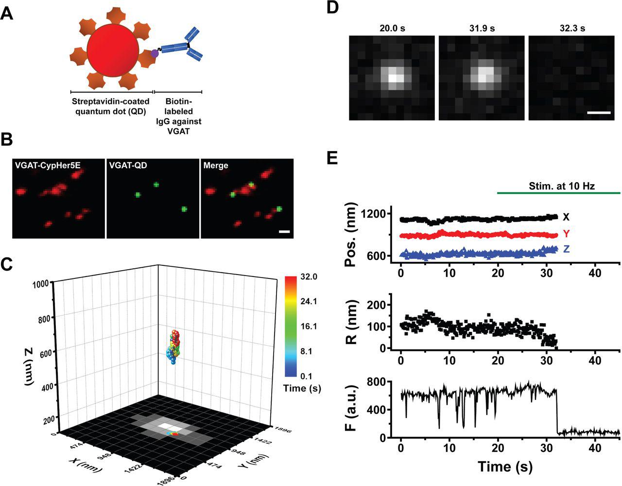 Streptavidin Conjugated Quantum Dots, supplied by Thermo Fisher, used in various techniques. Bioz Stars score: 99/100, based on 1 PubMed citations. ZERO BIAS - scores, article reviews, protocol conditions and more https://www.bioz.com/result/streptavidin conjugated quantum dots/product/Thermo Fisher Average 99 stars, based on 1 article reviews
streptavidin conjugated quantum dots - by Bioz Stars,
2026-06
99/100 stars
|
Buy from Supplier |
|
Carl Zeiss
confocal microscope carl zeiss inverted lsm 700 Confocal Microscope Carl Zeiss Inverted Lsm 700, supplied by Carl Zeiss, used in various techniques. Bioz Stars score: 90/100, based on 1 PubMed citations. ZERO BIAS - scores, article reviews, protocol conditions and more https://www.bioz.com/result/confocal microscope carl zeiss inverted lsm 700/product/Carl Zeiss Average 90 stars, based on 1 article reviews
confocal microscope carl zeiss inverted lsm 700 - by Bioz Stars,
2026-06
90/100 stars
|
Buy from Supplier |
|
Thermo Fisher
facs buffer  Facs Buffer, supplied by Thermo Fisher, used in various techniques. Bioz Stars score: 99/100, based on 1 PubMed citations. ZERO BIAS - scores, article reviews, protocol conditions and more https://www.bioz.com/result/facs buffer/product/Thermo Fisher Average 99 stars, based on 1 article reviews
facs buffer - by Bioz Stars,
2026-06
99/100 stars
|
Buy from Supplier |
|
Becton Dickinson
flow cytometry facscalibur Flow Cytometry Facscalibur, supplied by Becton Dickinson, used in various techniques. Bioz Stars score: 90/100, based on 1 PubMed citations. ZERO BIAS - scores, article reviews, protocol conditions and more https://www.bioz.com/result/flow cytometry facscalibur/product/Becton Dickinson Average 90 stars, based on 1 article reviews
flow cytometry facscalibur - by Bioz Stars,
2026-06
90/100 stars
|
Buy from Supplier |
|
Dilas Diode Laser
fluorescence imaging agent 17 Fluorescence Imaging Agent 17, supplied by Dilas Diode Laser, used in various techniques. Bioz Stars score: 90/100, based on 1 PubMed citations. ZERO BIAS - scores, article reviews, protocol conditions and more https://www.bioz.com/result/fluorescence imaging agent 17/product/Dilas Diode Laser Average 90 stars, based on 1 article reviews
fluorescence imaging agent 17 - by Bioz Stars,
2026-06
90/100 stars
|
Buy from Supplier |
|
Edinburgh Instruments
epl 475 ps pulsed diode laser Epl 475 Ps Pulsed Diode Laser, supplied by Edinburgh Instruments, used in various techniques. Bioz Stars score: 86/100, based on 1 PubMed citations. ZERO BIAS - scores, article reviews, protocol conditions and more https://www.bioz.com/result/epl 475 ps pulsed diode laser/product/Edinburgh Instruments Average 86 stars, based on 1 article reviews
epl 475 ps pulsed diode laser - by Bioz Stars,
2026-06
86/100 stars
|
Buy from Supplier |
|
Ocean NanoTech
green fluorescent quantum dots (qds Green Fluorescent Quantum Dots (Qds, supplied by Ocean NanoTech, used in various techniques. Bioz Stars score: 90/100, based on 1 PubMed citations. ZERO BIAS - scores, article reviews, protocol conditions and more https://www.bioz.com/result/green fluorescent quantum dots (qds/product/Ocean NanoTech Average 90 stars, based on 1 article reviews
green fluorescent quantum dots (qds - by Bioz Stars,
2026-06
90/100 stars
|
Buy from Supplier |
|
AnaSpec
fluorescent probes and quantum dots Fluorescent Probes And Quantum Dots, supplied by AnaSpec, used in various techniques. Bioz Stars score: 90/100, based on 1 PubMed citations. ZERO BIAS - scores, article reviews, protocol conditions and more https://www.bioz.com/result/fluorescent probes and quantum dots/product/AnaSpec Average 90 stars, based on 1 article reviews
fluorescent probes and quantum dots - by Bioz Stars,
2026-06
90/100 stars
|
Buy from Supplier |
Image Search Results
Journal: bioRxiv
Article Title: Inhibitory synaptic vesicles have unique dynamics and exocytosis properties
doi: 10.1101/2020.09.21.289314
Figure Lengend Snippet: (A) Schematic diagram depicting a streptavidin-conjugated quantum dot (QD) conjugated to biotinylated antibodies against the luminal domain of VGAT. (B) Colocalization of VGAT-QD‒loaded inhibitory vesicles (green) and CypHer5E-VGAT‒labeled presynaptic boutons (red) in cultured hippocampal neurons. Scale bar: 1 µm. (C) Three-dimensional trajectory of a VGAT-QD‒loaded inhibitory vesicle overlaid on the x - y plane of a CypHer5E-VGAT‒ labeled presynaptic bouton. The color bar represents elapsed time; electrical stimulation (10 Hz) started at 20 s, and the vesicle underwent exocytosis at 32.0 s. (D) Fluorescence images of the VGAT-QD‒loaded vesicle shown in panel C taken at the indicated times. Scale bar: 0.5 µm. (E) Three-dimensional position, radial distance from the momentary position to the fusion site (R), and fluorescence intensity (F) of the VGAT-QD‒loaded vesicle shown in panel C. Note the photoblinking events (e.g., at approximately 8 s, 13 s and 15 s), confirming the presence of one QD inside the vesicle. Electrical stimuli (10 Hz) were applied for 120 s starting at 20 s (green horizontal bar).
Article Snippet: The biotinylated monoclonal mouse anti-Syt1 antibody (105 311BT, Synaptic Systems) or the biotinylated anti-VGAT antibody (131 103CpH, Synaptic Systems) was conjugated to
Techniques: Cell Culture, Labeling, Fluorescence
Journal: iScience
Article Title: Preoperative immune checkpoint inhibition and cryoablation in early-stage breast cancer
doi: 10.1016/j.isci.2024.108880
Figure Lengend Snippet:
Article Snippet: Briefly, one million PBMCs and TILs were washed with 2 mL
Techniques: Recombinant, Saline, Staining, Extraction, Software, Flow Cytometry